Physical Description
External appearance
Dorsal:
Pseudobiceros bedfordi has a brownish-black body with uneven transverse pink streaks and numerous small yellow or white dots randomly dispersed across the entire dorsal surface ( Newman & Cannon, 1994).
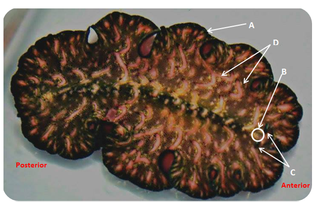
Figure 1. Dorsal anatomy of P. bedfordi. A. Body margin of flatworm lined with tiny epidermal mechanoreceptor cells. B. A cluster of multiple black dots (~100) known as eyespots form the cerebral eyespot. C. Pseudotentacles are formed from anterior foldings of the margin of theflatworm. D. Transverse pink streaks.
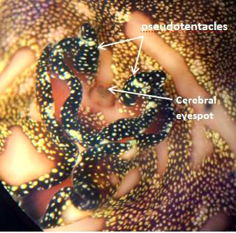
Figure 2. Dorsal surface: Close up view of pseudotentacles and cerebral eyespot in the head region of P. bedfordi.
Ventral view:
The ventral (bottom) surface is pink with a black band running around the perimeter of the body.
This is also where the mouth, genital pores (male and female) and an oral sucker is found.
Polyclads have their mouth positioned at the rear end. They feed by crawling over their prey and use their mouth apparatus to feed.The oral sucker may look small and insignificant , but they have a surprisingly strong, muscular pharynx that is able to crush and suck on prey.
Pseudobiceros is characterised by having 2 male reproductive systems (Faubel, 1984) hence possessing 2 male pores as seen in figure 2.
Figure 3. Ventral surface : Labelled regions of flatworm with pharynx located distinctly at anterior part of the flatworm.
Size
The adults range in size from 8- 10 cm in length.
Body Wall
The body surface is covered by a single layer of ciliated epidermis and each ciliated cell has many cilia (small, hair-like structures).They get the name "turbellaria" from the characteristic swirling motion generated by the cilia.
For in-depth description of cell structures forming the epidermis, head to " Anatomy and Physiology".
|